The Science
of XTi PULSE
XTi PULSE light therapy combines patented optical science with emerging bioscience to enhance human health. Here's how it works.
XTi PULSE light therapy combines patented optical science with emerging bioscience to enhance human health. Here's how it works.
XTi PULSE light therapy works by using light to improve how we feel, sleep, and physically and mentally perform. The sun is our inspiration. XTi PULSE light therapy activates the same natural pathways as sunlight and, when it’s time to sleep, the absence of light – which is hard to come by given the all the artificial light from the bulbs and screens of modern life.
Most of the light our eyes capture – and the darkness they also register – goes to parts of the brain that let us see. But some of it ends up in other parts of the brain that that influence and regulate sleep, inflammation, mood and behavior, cognitive performance, stress and anxiety, heart rate, respiratory rate, digestion, and more.
XTi PULSE therapy lights tap into that natural pathway with very specific light we’ve tested and refined for more than a decade, and that scientific work continues [link to current research below] with ongoing studies at the University of Colorado, Colorado State University, and others. XTi PULSE therapy light consists of combinations of colors in the visible spectrum (that is, light our eyes can see) and imperceptibly quick pulses within those colors.
We’ve patented these light combinations, which we call pulsed alternating wavelengths, or PAWs. Just as the sun and darkness have different effects on our minds and bodies, so do XTi PULSE’s different colors and pulses. One key difference: Our technology works much faster than natural sunlight, and we can tune it to achieve different impacts.
A key advantage of tapping into the brain’s natural light-related mechanisms is that it’s safe. XTi PULSE therapy light helps our bodies provide for themselves in achieving an optimal state, but the brain has dealt with light for eons, and it seems to know when enough is enough. Thousands of hours of use have shown no side effects. The light itself is gentle and safe for the eyes, and with no potentially harmful ultraviolet or infrared light.
So, that’s the big picture. For more detail, read on.
Our eyes are light sensors. They evolved mainly to improve the odds of finding or catching food—and reducing our odds of becoming food. So, it makes sense that most of the information passed from our eyes to our brains ends up in the visual cortex. There, the actual world around us, having been translated into electrical and chemical (photochemical) signals, gets reassembled into what we perceive to be the world around us.
The retina at the back of the eye captures incoming light and translates light into photochemical signals. We learned about the retina’s rods and cones in school—rods for low-light vision, cones for colors. But the retina’s many layers also host other cell types—among them horizontal cells, bipolar cells, amacrine cells, and, for our purposes most importantly, intrinsically photoactive retinal ganglion cells (ipRGCs). Discovered in 1998 in the African clawed frog and subsequently in mice, humans, and other mammals), they represent a third class of photoreceptor cells alongside rods and cones.
In the early 2000s, researchers showed that ipRGCs send photochemical signals to a part of the brain’s hypothalamus that controls circadian rhythms. It’s called the suprachiasmatic nucleus, or SCN. The circadian rhythms that the SCN regulates play an important role in our moods and all sorts of other biological functions. In addition, ipRGCs send signals to the perihabenular nucleus in the thalamus, which activates multiple brain regions, including the prefrontal cortex that’s responsible for high-level thinking such as problem solving.
The SCN activates other areas of the hypothalamus—a deep-brain structure that’s in charge of keeping the body in a stable state (homeostasis) on a variety of fronts. The hypothalamus regulates body temperature, emotions and behavior, the sleep-wake cycle, blood pressure, thirst, hunger, sex drive, and more. It does so by producing hormones that activate brain regions that control the autonomic nervous system.
Those retinal ipRGCs, via the SCN and the perihabenular nucleus, influence a wide range of neurological and physical activity. That’s this natural mechanism that XTi PULSE light therapy taps into and optimizes.
To understand how XTi PULSE light therapy’s pulsed alternative wavelengths (PAWs) do that, read on.
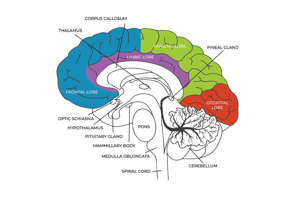
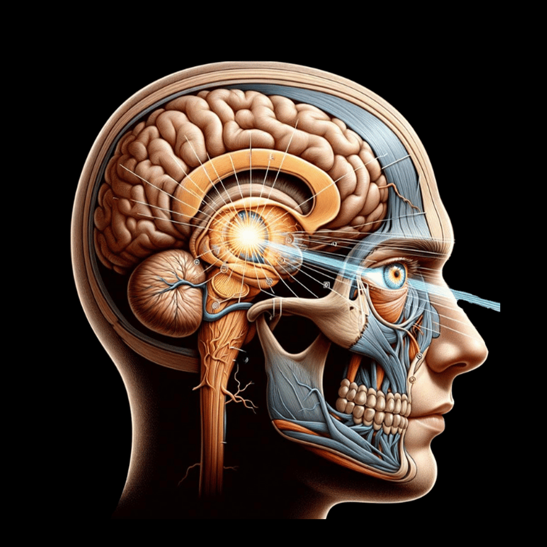
The retina’s ipRGC cells transmit photochemical signals not to the vision-producing occipital lobe in the brain’s posterior regions, but to the suprachiasmatic nucleus (SCN) in the center of the brain. It’s located just behind the optic shiasma – a.k.a. optic chiasm – depicted here. The SCN then triggers a variety of biological functions via the hypothalamus, pineal gland, and pituitary gland. The perihabenular nucleus in the thalamus also receives light signals from ipRGCs, in turn acting on various brain regions, including the prefrontal cortex – responsible for problem-solving – in the frontal lobe.
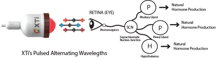
XTi PULSE light therapy’s patented pulsed alternating wavelengths (PAWs) recipes work by boosting the signaling from the retina’s intrinsically photoactive retinal ganglion cells (ipRGCs). These signals travel down the retino-hypothalamic tract (RHT) to the suprachiasmatic nucleus (SCN) and to the perihabenular nucleus. These brain regions, in turn, may activate other brain regions – depending, in part, on the nature of the light striking the retina and its ipRGCs.
XTi PULSE light therapy’s pulsed alternative wavelengths (PAWs) triggers a cascade of neurochemical reactions that influence a range of cognitive and physical function. This simplified view doesn’t include impacts via the thalamus’s perihabenular nucleus and the brain regions it affects.
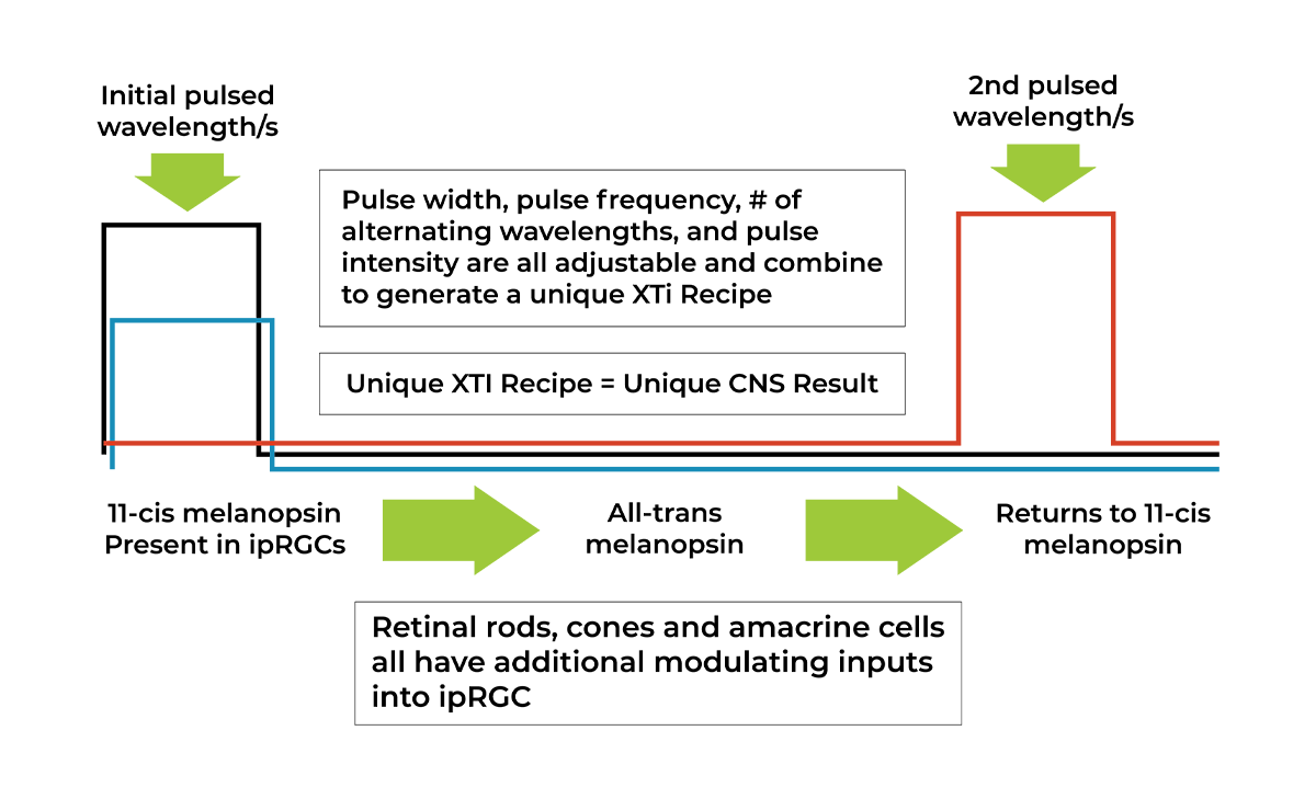
XTi PULSE light therapy’s main mechanism of action is through the protein melanopsin, the ipRGC’s primary photopigment. Melanopsin changes shape when hit by light, but it’s a slow-moving reaction, one that takes several seconds to complete. It’s even slower to recover, with a multistep chemical process dragging latency into the tens of seconds. That limits the pace of signaling to the SCN and perihabenular nucleus – and, with that, the positive impact of natural light on the brain (assuming there’s natural light available).
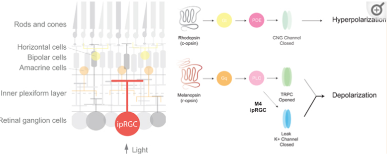
Rhodopsin is the critical protein in the highly light-sensitive retinal rods that let us see in very dim light. In ipRGCs, the key protein is the photopigment melanopsin. XTi PULSE light therapy works by drastically speeding up the signaling and recovery process of these proteins in the retina. That, in turn, boosts the delivery of beneficial neurotransmitters and hormones to various brain regions. Depending on the light, different brain regions have different chemical reactions. Hence the varying outcomes with XTi PULSE Performance and XTi PULSE Sleep & Recovery light-therapy recipes.
XTi PULSE light therapy uses pulses of light – PAWs – to speed up melanopsin’s pace of recovery and sharply reduce its latency. The first pulse turns 11-cis melanopsin into all-trans melanopsin, and that kicks off a neurochemical signal via the RHT. A second pulse provides the all-trans melanopsin the energy it needs return to its 11-cis melanopsin form. The process then repeats, thousands of times a second.
The SCN and the perihabenular nucleus receive much more frequent – and therefore more potent – signals than natural light can deliver, and they directly or indirectly influence a wide variety of physical and cognitive functions. One example: Data from animal-model experiments showed melatonin levels to leap by about 500% under one of our sleep recipes.
The XTi PULSE light therapy-induced photochemical process causes the photopigment protein melanopsin in ipRGCs to signal the brain and recover thousands of times faster than is possible with natural light – thus drastically strengthening the positive biochemical response.
Different PAWs wavelengths trigger different neurological outcomes and have varying biological effects, so different XTi PULSE light therapy recipes foster sleep & recovery and cognitive and physical performance. Health and wellness impacts appear to depend on the condition in question. For example, the XTi PULSE Performance recipe improves focus and concentration for ADHD patients and neurotypical users alike. The XTi PULSE Sleep & Recovery recipe diminishes anxiety, tempers Parkinson’s disease symptoms, and helps people sleep, among other observed effects.
The precise biochemical roots of those effects are still largely unclear. Why? Consider that the suprachiasmatic nucleus alone influences the production of melatonin, cortisol, leptin, ghrelin, growth hormone, and thyroid stimulating hormone, with diverse effects.
Add to all that the complexities of the perihabenular nucleus. It’s part of the lateral habenula that acts as a hub integrating value-based, sensory, and experiential information to regulate diverse motivational, cognitive and motor processes.
Suffice it to say that XTi PULSE light therapy taps into extraordinarily complex, phylogenetically ancient, undeniably potent neurochemical processes. XTi PULSE light therapy’s impact on sleep, recovery, performance, and health and wellness is far easier to observe than its modes of action, which remain an area of great interest in neuroscientific research.
Our research program launched in 2014, when the first of our more than 75 patents and pending patents was issued. Based in the agricultural hub of Greeley, Colorado, we at first focused on PAWs’ observed positive impact on plant and crop growth, when then held true in studies of behavioral and health improvement in poultry and cattle. Starting in 2017, we set out to more systematically understand the sleep, performance, and human health impacts.
Our other research partners include:
This statement has not been evaluated by the Food and Drug Administration. This product is not intended to diagnose, treat, cure, or prevent any disease.